Dataset
This project uses the public Brain Tumor MRI Dataset from Kaggle (Masoud Nickparvar), which contains 7,023 MRI images across four classes: glioma, meningioma, pituitary, and no tumor.
Before training, I removed 218 duplicate images (191 training, 27 test) using MD5 hashing, leaving 5,521 training images and 1,284 test images.
Python Code
Purpose
This project aims to classify brain MRI scans into four classes and explain what the model looks at when it makes a prediction. The goal is a model that is both accurate and inspectable, since a black-box prediction is hard to trust in a clinical setting.
Method
- Preprocessing: resized images to 224x224, applied ImageNet normalization, and augmented the training set with horizontal flips, 10 degree rotations, and color jitter.
- Model: fine-tuned a ResNet50 pretrained on ImageNet (PyTorch). All layers were frozen except the last residual block (layer4), with a new classification head (2048 to 256, ReLU, Dropout 0.4, 4 outputs).
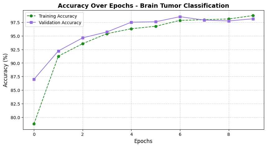
- Training: cross-entropy loss, Adam with weight decay, a step learning-rate schedule, and early stopping. Trained on a Colab T4 GPU; training stopped after 10 epochs.
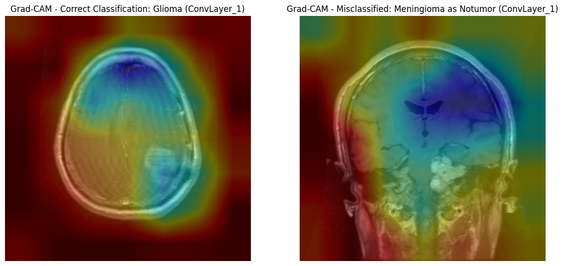
- Explainability: applied Grad-CAM and LIME to see which regions of the scan drove each prediction.
Results
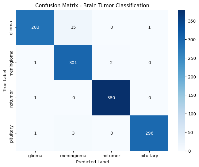
The model reached 98% accuracy and 0.98 macro F1 on 1,284 test images.
| Class | Precision | Recall | F1 |
|---|---|---|---|
| Glioma | 0.99 | 0.95 | 0.97 |
| Meningioma | 0.94 | 0.99 | 0.97 |
| No tumor | 0.99 | 1.00 | 1.00 |
| Pituitary | 1.00 | 0.99 | 0.99 |
Grad-CAM showed that the model sometimes attended to surrounding tissue rather than the tumor itself, which is a useful check on what the accuracy number does and does not mean.
Notes
The test set was also used for early stopping, so the 98% figure is not a fully independent holdout result. A separate validation split would be the next improvement.
Course: CSC 578 Neural Networks and Deep Learning, DePaul University (Winter 2025).